Structures & workspace
Open a structure. Start exploring.
Type an identifier or bring your own file. The structure arrives sorted into layers, ready to turn, style and measure, in your browser.
On screen, human hemoglobin (PDB 4HHB):
- 4 chains
- 574 residues
- 4 heme groups
Open
From an identifier to a structure on screen.
Type a PDB identifier, or bring your own file. The structure opens in the browser, with nothing to install, ready to turn and inspect.
Load by identifier or by file
A PDB identifier in the box at the top, or your own PDB, mmCIF, MOL2 or SDF file.
Turn, zoom and focus
Drag to rotate, scroll to zoom, and keep the whole structure in view or go in close.
Structures and their hierarchy
On the left, every structure with its chains, residues, ligands and waters.
Choose what a click selects
An atom, a residue, a chain or a whole entity.
Layers
Layers, each with its own style and colour.
A structure arrives sorted by kind of molecule. Each layer is shown, hidden, drawn and coloured on its own, so the picture says what you need it to.
Sorted on arrival
Protein, ligands, waters, ions and metals are separate layers from the start.
Three families of representation
Ribbon, atoms and bonds, and surface. Add more than one to the same layer.
Colour by what matters
By chain, element, residue, secondary structure, hydrophobicity or B-factor, or in a colour of your own.
Opacity and size
Set for each representation, so one can step back while another comes forward.
Measure
Measure what matters.
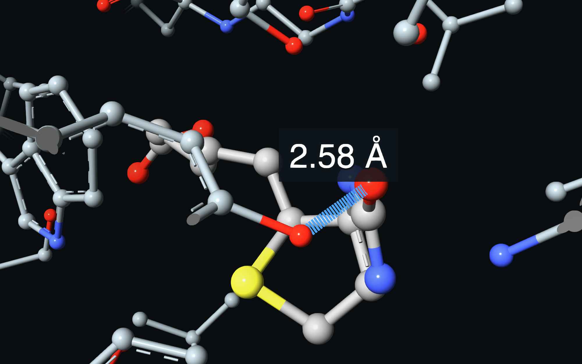
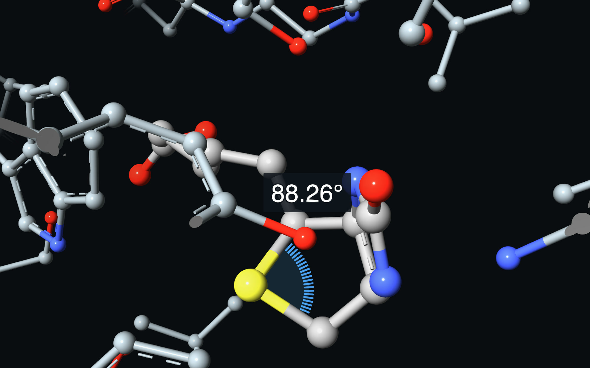
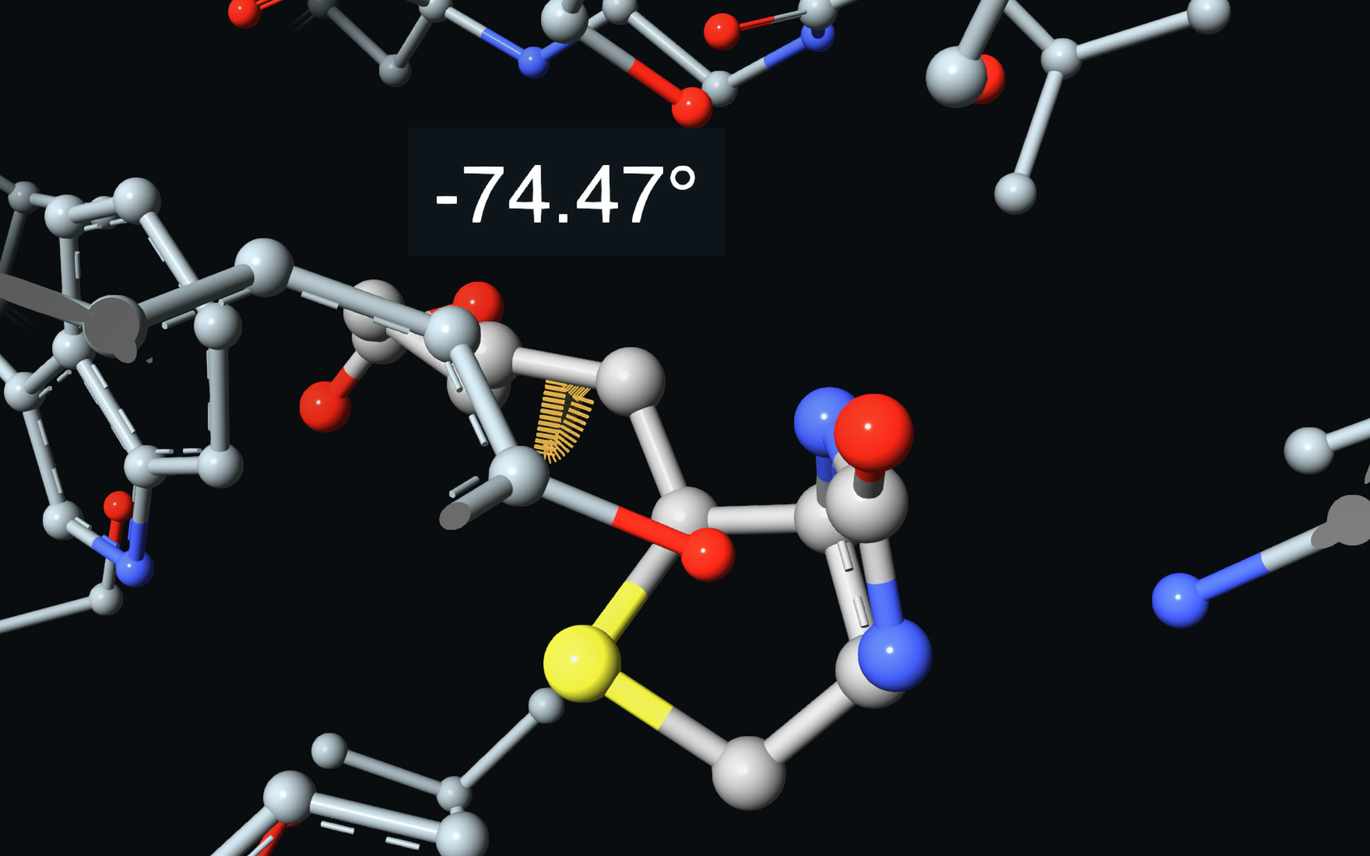
Pick atoms on the structure and read the geometry between them. Every measurement stays on screen and in a table. Here, on biotin in its binding site.
Distances
Between two atoms, drawn as a dashed line with its value in ångström.
Angles
Between three atoms; the second one is the vertex.
Dihedrals
Four atoms in order, for the twist around the bond between the middle two.
A table of measurements
Review the values and the atoms they were taken from, and remove the ones you no longer need.
Beyond proteins
Every kind of molecule, drawn its own way.
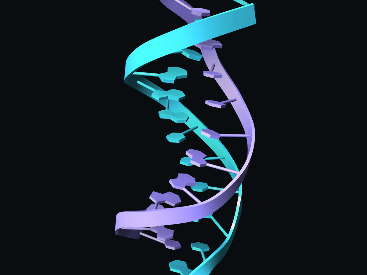
Nucleic acids
DNA and RNA with base rings or base blocks, or the backbone alone.
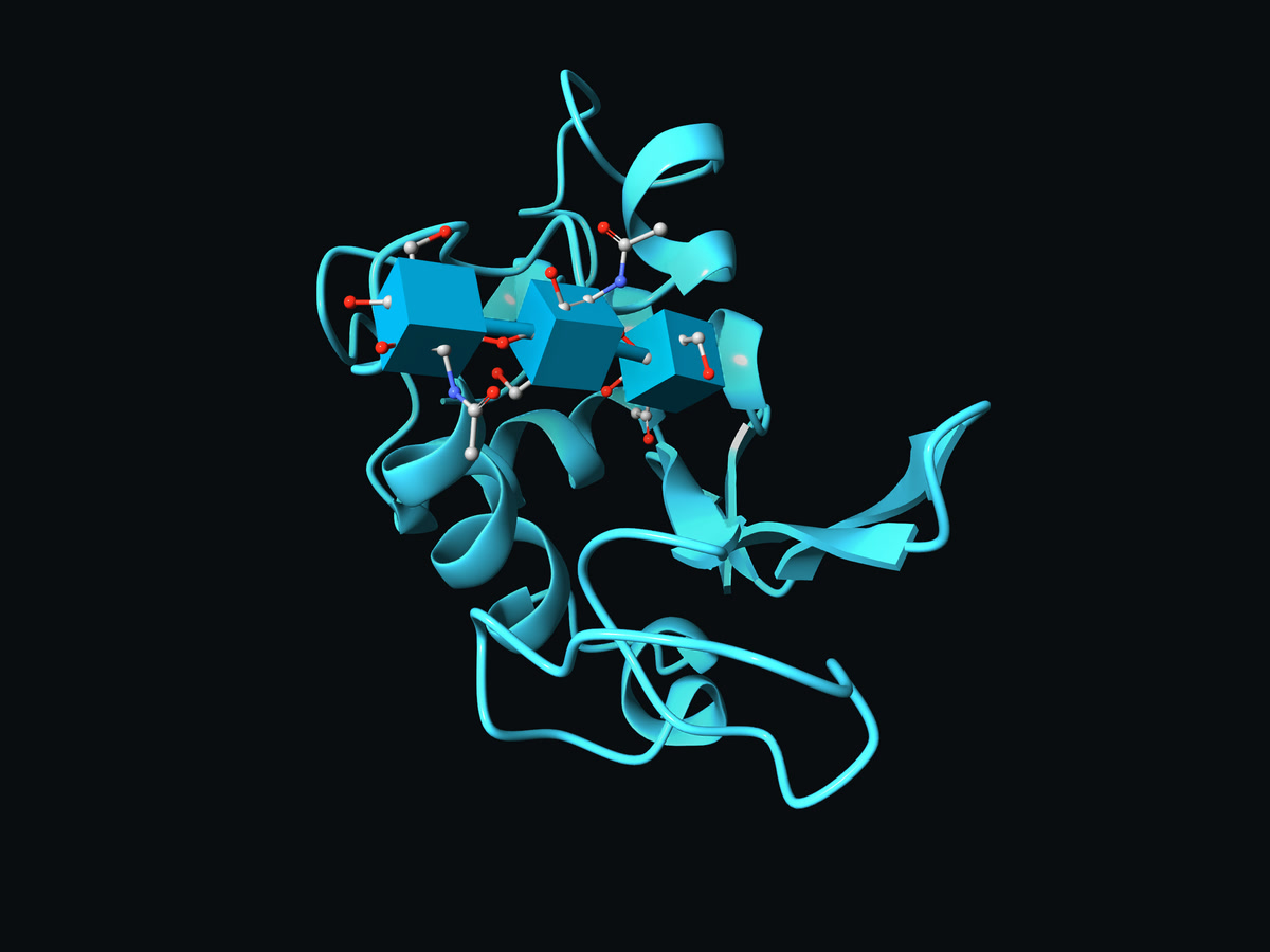
Carbohydrates
Sugars as their standard symbols, with or without their atoms.
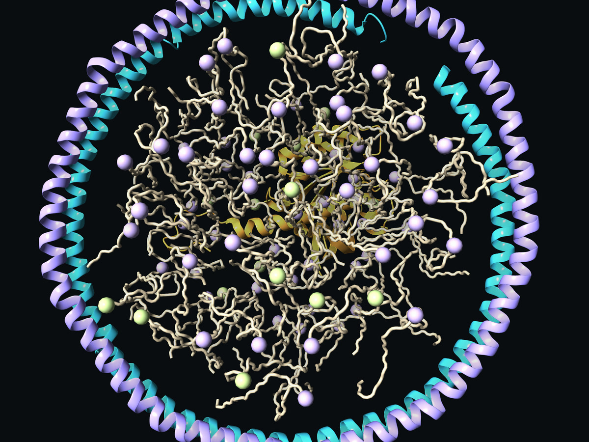
Lipids
Head groups and tails drawn apart, to read a membrane at a glance.
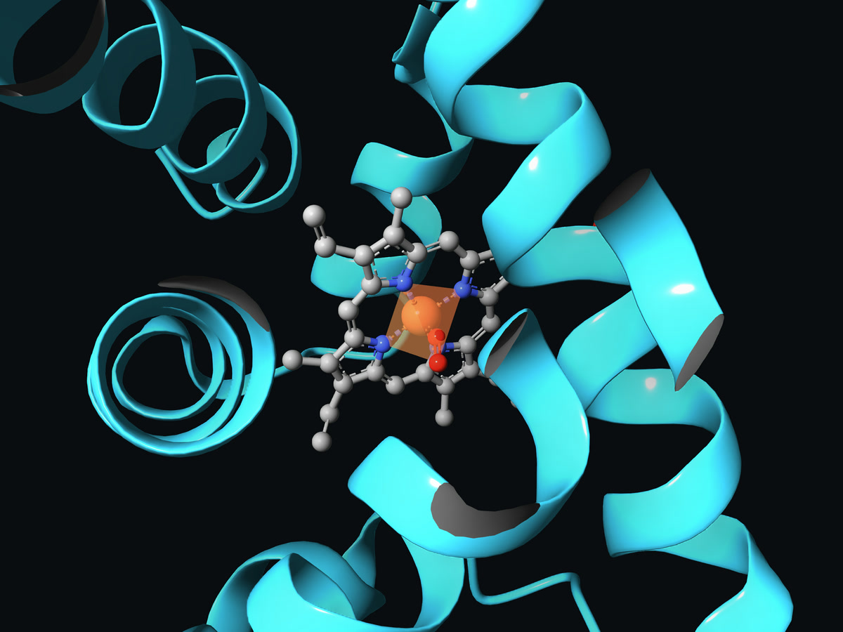
Metals
Found when the structure loads, with their coordination geometry when you ask for it.
Next